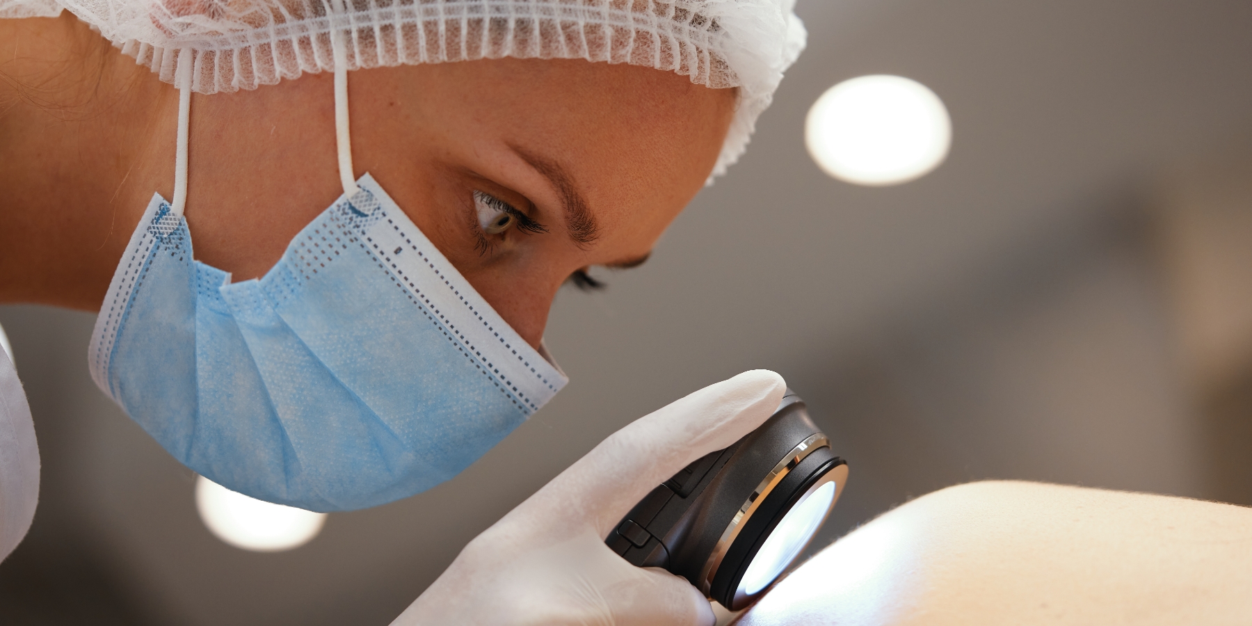
Skin cancer affects millions of Americans annually, but early detection and treatment significantly improve outcomes. Mohs surgery is a highly effective, tissue-sparing technique that removes cancerous cells while preserving healthy skin, making it an ideal choice for sensitive areas like the face and hands. At Houston Skin, we’re committed to offering this advanced procedure to our patients, providing the most precise care and optimal results possible.
What Is Mohs Surgery?
Mohs micrographic surgery is an advanced surgical technique designed specifically for the removal of skin cancers. Named after Dr. Frederic Mohs, the inventor of the technique, this procedure is based on the principle of using a microscope to trace skin cancer roots so that the cancer may be completely removed. This technique has the highest cure rate for the most common types of skin cancer: basal cell carcinoma and squamous cell carcinoma. The entire tumor is removed with the smallest possible defect, potentially resulting in less scarring at the site. At Houston Skin, our priority is to deliver quality care, information, and treatment for Mohs surgery in a convenient, welcoming environment.
Will My Insurance Cover Mohs Surgery?
Most health insurance plans will cover the costs associated with Mohs surgery and any other wound repair treatments. However, all insurance plans are different, and it is always important to verify the specifics of your plan. Our team is happy to answer any questions you have about the insurance claims process.
What To Expect
During Mohs micrographic surgery, your Mohs surgeon removes a piece of tissue just around the visible tumor on the skin surface. This tissue is examined with a microscope to identify any cancerous roots that extend beyond the visible boundaries of the tumor. If any remaining cancerous cells are identified by microscope, your Mohs surgeon will return to the remaining tumor cells and remove another layer of tissue.
This tissue will then be examined for any additional cancer cells. This process continues until no further cancerous cells are identified. Taking the tissue in layers and examining each with the microscope ensures that all the cancerous tissue is removed and prevents the unnecessary removal of healthy tissue.
Once the tumor has been removed, reconstructive surgery is typically performed to repair the resulting defect. This surgery typically requires the placement of sutures to achieve the optimal cosmetic appearance.
Recovery After Mohs Surgery
Following your Mohs surgery, it will be important to care for the treatment site by regularly changing your bandages. Your dermatologist will provide full instructions before you are discharged, and our Houston Skin team is always available to answer questions by phone.
Mohs Surgery FAQs
-
Will There Be Pain After Surgery?
Some patients experience mild discomfort in the days following their Mohs surgery. These symptoms can usually be managed with over-the-counter medications like Tylenol®. It is important to avoid any aspirin-derived products, including Advil®, as these products can increase your risk of bleeding.
-
Will Mohs Surgery Leave a Scar?
While some degree of scarring is expected with any surgical procedure, our team at Houston Skin is dedicated to providing meticulous cosmetic closures. We understand that the appearance of your skin matters, and we employ advanced techniques to minimize scarring and optimize the aesthetic outcome of your treatment. You can trust us to prioritize both your health and your confidence.
-
Are There Any Complications?
Mohs surgery is known for being safe and effective, and most patients tolerate it well. However, there can be risks with any surgery. Possible complications from Mohs surgery include bleeding, bruising, infection, physical discomfort, scarring, and poor wound healing.
Meet The Team
Our Houston team of Board-Certified Dermatologists and licensed skin care providers brings a wealth of experience and a passion for healthy skin. With extensive clinical experience and the highest levels of training, our doctors are committed to delivering exceptional care and achieving optimal results for every patient. We prioritize your comfort and safety, promoting a positive and reassuring experience throughout your journey to vibrant skin. We’re honored to be your trusted provider of quality medical dermatology care and your partners in achieving and maintaining your skin’s health.
Contact Houston Skin for Mohs Surgery
Schedule a Mohs surgery consultation at one of our convenient Houston Skin locations today. Our board-certified dermatologists will provide a personalized assessment and recommend the ideal treatments to help you heal and remove skin cancer. Call now or schedule a consultation online to take the first step towards a more confident, radiant you.


